Regenerative Medicine
Alopecia and Hair Thinning: The Power of Regenerated Follicles
Those who notice their brush fuller than usual, or a parting that widens month after month, arrive at their visit with a single question: can something still be done? The answer almost always depends on the condition of the follicle, much more than on the quantity of hair already lost.
At IMAGE REGENERATIVE, at our Milano and St. Moritz locations, pathways dedicated to alopecia and hair thinning start exactly here, by understanding whether the follicle is still vital and how much biological margin remains to reactivate it.
The hair follicle is a living structure, capable of going through resting phases and reactivating even after years of suffering. On this plasticity is based the regenerative medicine applied to the scalp, which addresses thinning by working on the environment in which the follicle lives even before the visible hair.
Alopecia and hair thinning: what they are
Thinning and alopecia describe two moments of the same process. Thinning is a progressive rarefaction, in which hair remains but becomes thinner, shorter and less pigmented, until the scalp begins to show through in backlight. We speak of alopecia when the loss becomes evident and leaves uncovered areas, with a pattern that varies depending on the clinical form.
At the base is the hair cycle, which each follicle repeats throughout life. The anagen phase is that of active growth and lasts from two to six years. The catagen phase is a brief transition, of a few weeks, in which activity stops. The telogen phase is rest, after which the hair falls out to make room for the new one. In a balanced scalp the vast majority of follicles are in anagen, and losing up to a hundred hairs a day is within normal range.
The problem arises when this ratio shifts. If the growth phase shortens and the resting phase lengthens, each cycle returns a hair slightly smaller than the previous one. This is the phenomenon of miniaturization, the true engine of progressive thinning, and explains why the perception of less full hair precedes by far the appearance of truly uncovered areas.
Androgenetic alopecia and alopecia areata: the differences
The two most frequent forms have opposite mechanisms and different prognoses, so distinguishing them is the first useful step.
Androgenetic alopecia is the most common overall and affects most men and about one in two women over the course of their lives. It depends on the combination of hereditary predisposition and follicle sensitivity to androgens. It has a slow course and a recognizable pattern. In men the frontal hairline recedes and the vertex thins, in women the central parting widens while the front hairline generally remains preserved.
Alopecia areata has an autoimmune origin, because the immune system attacks the follicle in the growth phase and blocks its activity. It appears rapidly, with roundish patches with sharp margins, often in a single area of the scalp or beard. Unlike the androgenetic form, the follicle remains structurally intact, and for this reason spontaneous regrowth is frequent, although the disorder tends to reappear over time.
There is also a third group, scarring alopecias, in which inflammation replaces the follicle with fibrous tissue. Here the follicle is lost irreversibly, and recognizing them early radically changes the available options.
The causes: hormones, stress and predisposition
In clinical practice the factors that sustain hair loss almost always intertwine, but four recur more than others:
· Dihydrotestosterone, derived from testosterone by the action of the enzyme 5-alpha-reductase, shortens the growth phase and thins the shaft with each successive cycle.
· Genetic predisposition, polygenic and inherited from both family branches, establishes how sensitive individual follicles are to that same hormone.
· Intense physical or emotional stress pushes a quota of hair early into the resting phase, with abundant shedding that manifests two to three months after the triggering event.
· Nutritional deficiencies and certain medical conditions, from iron deficiency anemia to thyroid dysfunctions to certain drug therapies, weaken the follicle in a diffuse and reversible way.
To these are added elements that rarely act alone but worsen the picture, such as smoking, severely restrictive diets, prolonged sun exposure without protection and aggressive cosmetic treatments repeated over time. In our team's experience, the most useful part of the first visit consists precisely in separating the stable component, linked to genetics and hormones, from the transitory and correctable one, because the two respond to completely different strategies.
Female alopecia: specificities and causes
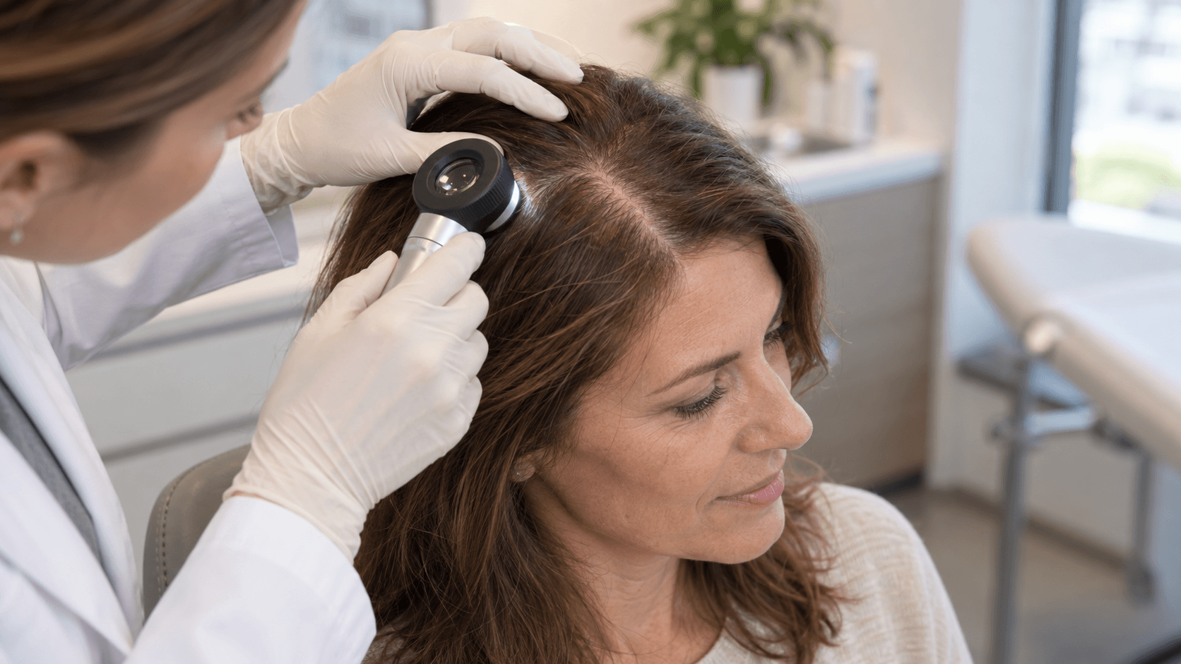
In women, hair loss follows its own rules and is recognized later, because the pattern is less apparent. The initial sign is almost always the widening of the central parting, accompanied by the sensation of a thinner ponytail at the base. The frontal hairline, on the other hand, tends to remain intact even when thinning in the central area is already advanced.
The hormonal phases of female life punctuate the phenomenon. After childbirth, the collapse of estrogens causes a physiological telogen effluvium that resolves within a few months. In menopause, on the contrary, the reduction of estrogenic protection is stable and allows the androgenetic sensitivity of follicles to emerge, with thinning that progresses slowly but without spontaneous reversals. Polycystic ovary syndrome and thyroid dysfunctions represent the other two conditions to systematically exclude.
An aspect that deserves attention is the overlap of pictures, because many patients simultaneously present an underlying androgenetic component and a transitory effluvium triggered by a diet, a surgical intervention or a period of stress. Treating only one or only the other leads to partial results, and trichoscopy associated with a targeted blood profile serves exactly to establish which of the two components weighs more.
The power of the regenerated follicle: regenerative medicine for hair
A miniaturized follicle is profoundly different from a lost follicle. In the first case the structure is still present, the niche of stem cells is preserved and the tissue maintains the capacity to respond to an adequate stimulus. In the second, fibrosis has replaced the follicular unit and no biological therapy can reconstruct it. All regenerative medicine applied to hair works within this margin.
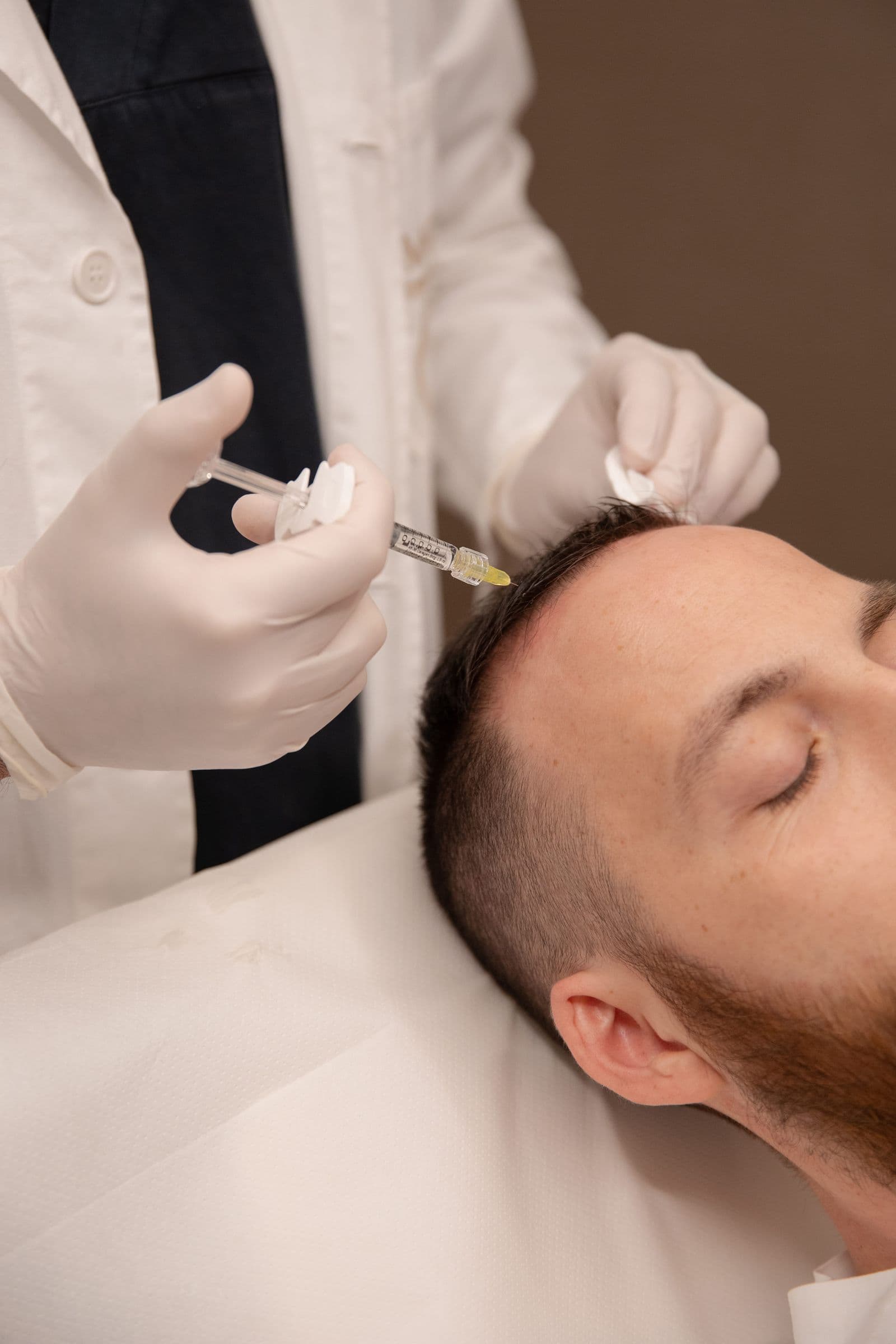
The objective is to modify the environment in which the follicle lives, thus improving microcirculation, favoring neoangiogenesis, reducing the inflammatory state of the scalp and providing fibroblasts with signals to reconstruct a support matrix. Hair filler acts on this level through micro-injections of biomimetic peptides delivered by hyaluronic acid, with a protocol of three or four sessions at intervals of three to four weeks and maintenance sessions at intervals of six to twelve months.
On the more properly regenerative side, autologous micro-fragmented adipose tissue with the Lipogems® method, developed by Prof. Carlo Tremolada, brings stromal cells, growth factors and intact extracellular matrix to the scalp. However, an honest clarification is needed. These approaches support a weakened follicle, while in the face of advanced baldness the correct indication remains another. The clinical reasoning behind these choices is also collected in our in-depth study on hair thinning and loss.
Comparing treatments: PRP, drugs and regenerative therapies
The choice between available options depends less on their theoretical efficacy and more on where the patient stands. Topical and oral drugs remain the reference standard for slowing the androgenetic component, with a limit that must be stated clearly, because they act as long as they are taken and upon discontinuation the process resumes its course.
Platelet-rich plasma deserves a clarification, because it is often presented as an alternative. In our practice it is a complementary treatment, which releases growth factors in the scalp and pairs well with both injectable protocols and surgery, without replacing one or the other.
Hair transplant comes into play when miniaturization has passed the point of no return. The microsurgical technique performed without shaving harvests follicular units from the occipital area, genetically resistant to androgens, and repositions them among existing hair. Visible growth begins around the fourth month and continues for the following year.
Approach | How it works | What it requires over time |
Topical or oral drugs | Slow miniaturization by acting on hormone or microcirculation | Continuous intake: upon discontinuation the picture progresses again |
PRP | Autologous platelet concentrate that releases growth factors in the scalp | Repeated cycles; integrates with other treatments rather than replacing them |
Hair filler | Biomimetic peptides and hyaluronic acid for microcirculation and follicular vitality | Three or four close sessions, then six-monthly or annual maintenance |
Regenerative transplant | Repositions follicular units resistant to androgens, without shaving | Single repeatable session; complete result within twelve months |
How to recognize and when to intervene

Some signs anticipate by months the perception of less full hair, such as hair that breaks more easily, a ponytail or bun visibly less voluminous with the same gathering, a widening parting, scalp that shows through in backlight and abundant and prolonged shedding beyond four to five weeks. Taken individually they are worth little, together they indicate that the cycle has already shifted.
The clinical evaluation relies on trichoscopy, which measures density, shaft diameter and proportion between thin and terminal follicles, and a targeted blood profile on ferritin, thyroid function, vitamin D and hormonal status. On these two elements depends the distinction between reversible shedding and structured miniaturization.
On when to intervene the answer is less negotiable than one might think, because the margin for recovery coincides with the number of still vital follicles and that number decreases over time. Anticipating the first evaluation when doubt appears, instead of waiting for thinning to become evident, changes the available options and extends the therapeutic horizon by years. The Milano and St. Moritz locations follow the same initial protocol, with the possibility of immediately setting up a pathway for improving hair quality.
Frequently asked questions
Can the hair follicle be regenerated?
A miniaturized follicle preserves its niche of stem cells and can return to producing thicker hair if the surrounding environment improves. A follicle replaced by fibrous tissue, on the other hand, is permanently lost: trichoscopy serves precisely to distinguish the two situations before setting up any therapy.
How do you distinguish androgenetic alopecia from alopecia areata?
Androgenetic has a slow course and a pattern design, with progressive thinning of hair in areas sensitive to androgens. Areata appears rapidly, with roundish patches with sharp margins and smooth scalp, and has an autoimmune origin. The prognosis is also different: in areata spontaneous regrowth is frequent.
Is female thinning reversible?
It depends on the cause. A post-partum telogen effluvium or one linked to iron deficiency resolves when the triggering factor is corrected. The androgenetic component, on the other hand, tends to progress and is managed over time with maintenance protocols. In most patients the two components coexist.
When is it too late to intervene?
The criterion is not age but the vitality of residual follicles. As long as trichoscopy shows a significant quota of miniaturized hairs, there is margin for regenerative therapies. When the area is devoid of follicular units, the appropriate option becomes transplantation.
Related Articles
 regenerative-medicine
Intravenous NAD+ Therapy: What It Is and What It's For
Intravenous NAD+ therapy: what it is, what it's for, and the benefits for energy and anti-aging. How the session works, differences with supplements, and side effects.
 regenerative-medicine
Deflated Breasts After Breastfeeding: Causes and Solutions
Deflated breasts after breastfeeding: causes and solutions, from exercises to firm the breasts to medical solutions like mastopexy and lipofilling. Complete guide.
 regenerative-medicine
Longevity, Anti-Aging and Biohacking: What They Really Mean
Longevity, anti-aging and biohacking: what they really mean and how they differ, between longevity medicine and optimization of body and mind.
Want More Info?
If you need more information, call us at +39 02 76280736, message us on WhatsApp, email us at info.milano@imageregenerative.com, or use the form below.